Open tresD DICOM viewer

Your CBCT, open in the browser.
Nothing to install. Free.

tresD DICOM viewer opens DICOM and DICOMDIR files and intraoral scans, and in seconds you can move through the MPR slices and see the 3D render, the panoramic, the TMJ, the airway and the superimposition of two studies. Research and teaching software.

Open tresD DICOM viewer

Your files are never uploaded: everything is processed on your own computer.

Created by Dr Manuel Fernández Cano, orthodontist
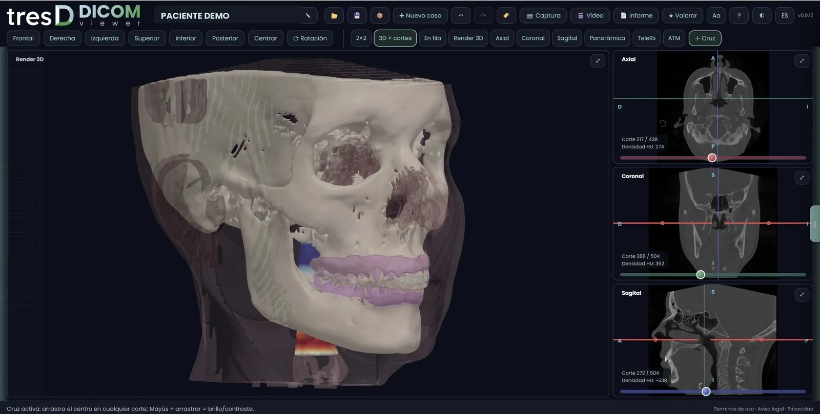
tresD DICOM interface with the 3D render and the three slices
100% in your browserChrome, Edge, Firefox or Safari, on Windows and Mac
Free, no accountNo sign-up needed to use it
Spanish, English, Portuguese and RussianSwitch language with one click
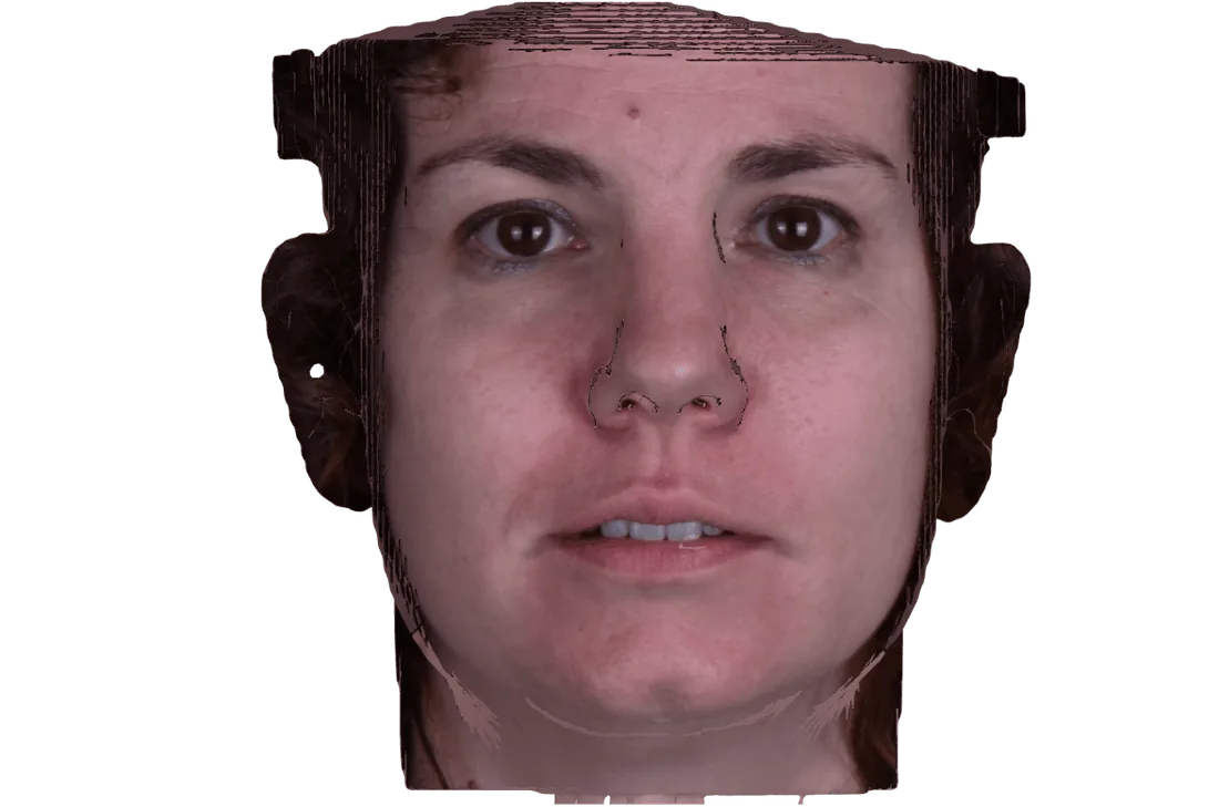
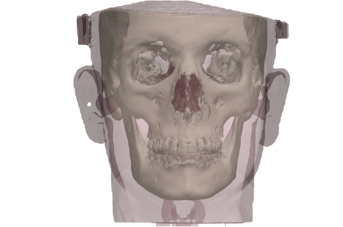
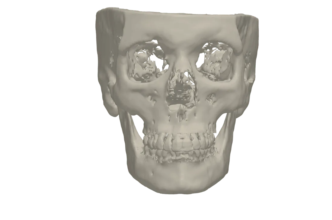
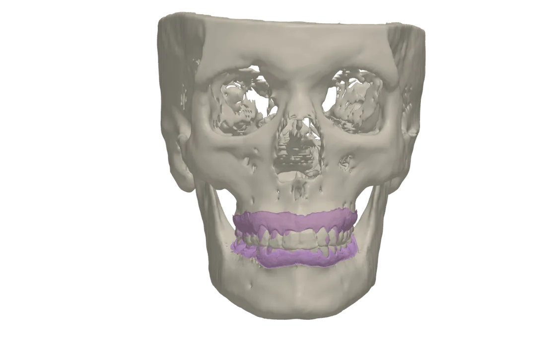
Patient photo draped over the skin reconstructed from the CBCT The skin turns transparent and the bone appears Bone segmented from the CBCT Intraoral scans placed on the bone Segmented airway with heat map

This is your patient

Builds a 3D soft-tissue file from a single photo

Scroll and the skin turns transparent

Soft tissue and bone, each with its own transparency.

The bone, segmented in seconds

automatically, with no thresholds to fiddle with.

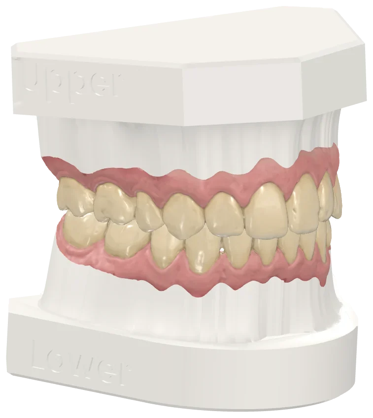
This is how it looks with the intraoral scans

STL, PLY or OBJ placed automatically on the bone.

And the airway

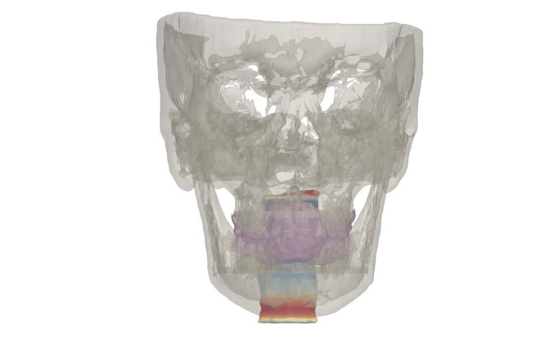
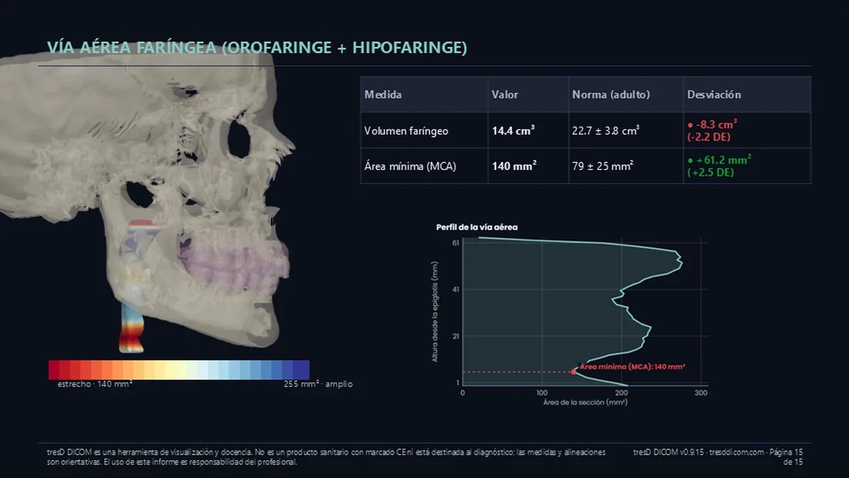
Volume, minimum area and a heat map by cross-section.

See it in action

In a few seconds: open the case, the 3 MPR slices, the 3D, the cephalograms and the panoramic.

Enjoy every one of its features

Get everything out of the CBCT

Superimposition of two studies

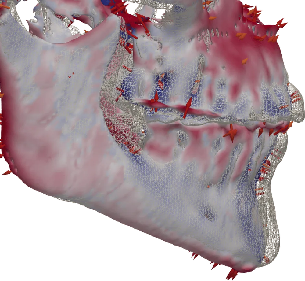
Load the initial and the final CBCT and tresD DICOM viewer superimposes them automatically, with no landmarks. A colour map shows how much each area moved and in which direction.

  • Automatic alignment on the cranial base
  • Colour map of differences in millimetres
  • Displacement arrows and T0 as a wireframe view
  • Works across CBCTs from different scanners (CT and CBCT)

How the superimposition works →

Superimposition of two CBCTs with colour map and arrows

Automatic panoramic

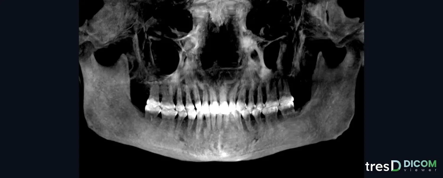
A panoramic taken from the CBCT itself, with a hand-editable arch curve and the thickness you choose, up to the condyles.

  • Curve drawn or corrected by hand
  • Browse the individual slices of every tooth

All about the panoramic →

Panoramic reconstructed from the CBCT

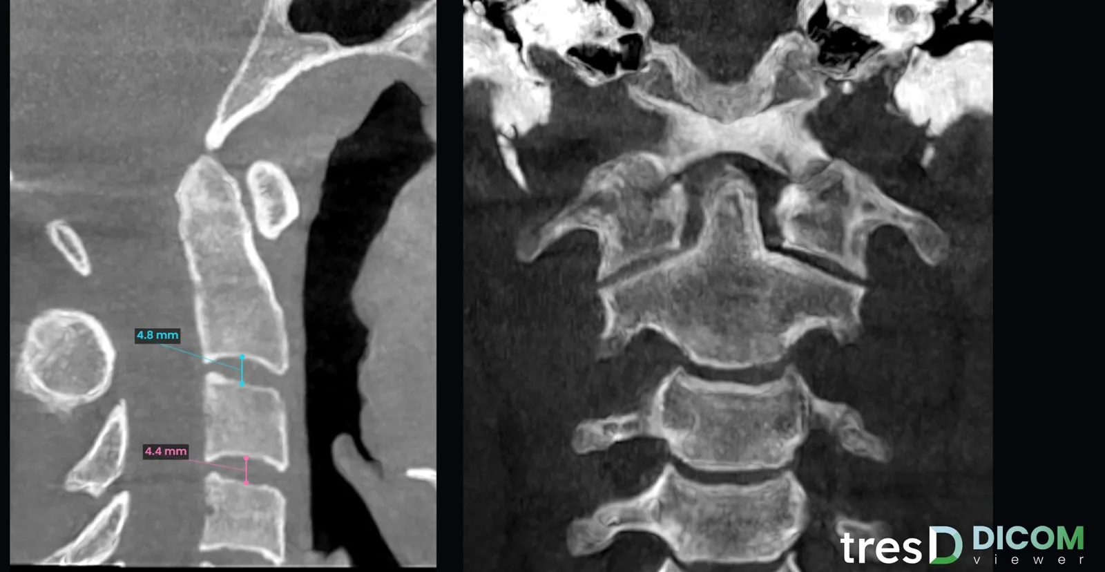
Cervical vertebrae in sagittal and coronal

A dedicated view with the sagittal and coronal slices of the cervical vertebrae, located automatically behind the airway. The wheel scrolls through the slices and the middle button adjusts the CBCT area.

  • Adjustable thickness and MIP
  • Indicative distance and angle measurements
  • Image filters
  • Screenshot with both slices, and in the report

Vertebrae, transverse slice and suture →

Sagittal and coronal slices of the cervical vertebrae with measurements

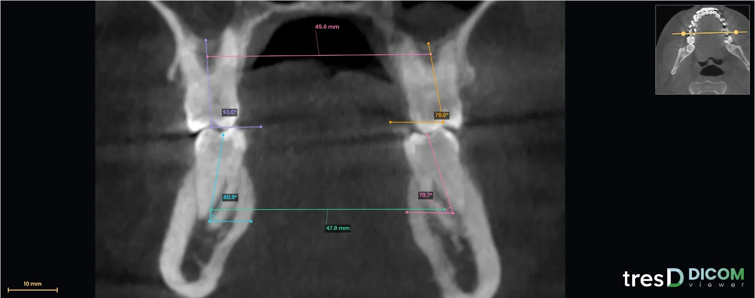
Transverse slice through the first molars

A high-resolution coronal slice through the first molars. Rotate and move it from the axial thumbnail, and use the wheel to move it forwards or backwards.

  • Axial thumbnail with the slice line
  • Indicative distance and angle measurements
  • Thickness, MIP and filters

More about the transverse slice →

Transverse slice through the first molars with measurements and axial thumbnail

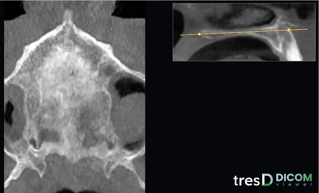
Midpalatal suture

A slice parallel to the ANS-PNS plane, found automatically, halfway through the palate thickness. Rotate it from the sagittal thumbnail and adjust the height with a slider.

  • Automatic ANS-PNS plane
  • Adjustable height and rotation
  • Thickness, MIP, filters and indicative measurements

More about the palatal suture →

Midpalatal suture slice with the sagittal thumbnail of the ANS-PNS plane

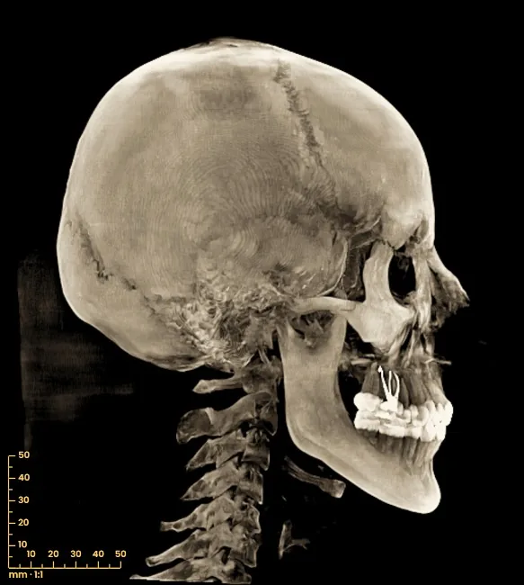
Lateral and frontal cephalograms, taken automatically from the CBCT

From the same CBCT you get the 2D lateral and frontal cephalograms, projected with parallel rays at 1:1 scale, in MIP or radiograph mode. No extra radiation to obtain the cephalogram.

  • Lateral and frontal, MIP or radiograph
  • 1:1 scale with a 5 mm ruler and adjustable tilt
  • Filters: hyperrealistic, sharp, soft, high contrast, inverted and ivory
  • Export your capture for cephalometric studies

All about the cephalogram →

Lateral cephalogram from the CBCT with the ivory filter

TMJ slices, right and left condyle

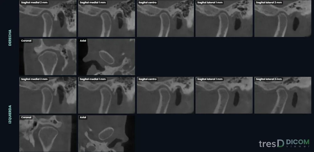
One click on each condyle and you get the sagittal, coronal and axial slices perpendicular to its axis, 1 mm apart, scrollable and with distance and angle measurements.

  • Both condyles side by side to compare them
  • Automatic framing on the joint
  • Indicative measurements on the slice

All about the TMJ slices →

TMJ slice mosaic of both condyles

Generate and download a report in seconds

Screenshots, measurements and notes go into a PDF or PowerPoint report. And the whole case is saved in one file so you can reopen it exactly as you left it.

  • PDF report and editable PowerPoint
  • You can pick the .tresd session back up with everything you did
  • .tresdz package to share the case with your colleagues
Airway page of the report with the table, the norm and the area profile

Each feature, step by step: Panoramic · Airway · TMJ · Superimposition · Cephalogram · Intraoral scan · DICOM to STL · Suture, transverse and vertebrae · iPad and phone · All

Video tutorials

17 short step-by-step videos to get the most out of it, from the first CBCT to the report. The videos are in Spanish: turn on YouTube subtitles and auto-translate them.

See the full playlist on YouTube

tresD Models: models with ABO base and engraved labels From the makers of tresD DICOM Do you also work with intraoral scans? Try tresD Models, our free orthodontic models program, also in the browser with nothing to install: realistic colour on your STLs, scan editing, measurements, virtual model bases ready to print and T0 – T1 superimposition. Try tresD Models for free
At our courses

tresD DICOM in the hands of the attendees

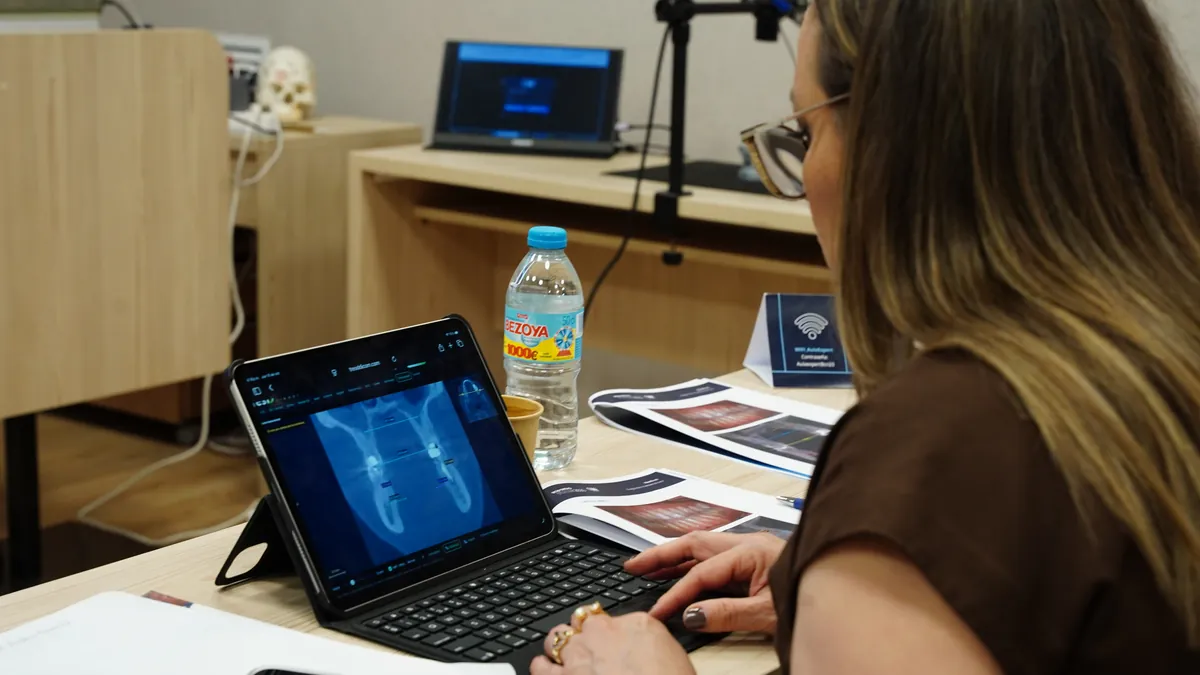
At our in-person courses, every attendee works with tresD DICOM on their own computer or tablet, in the browser.

An attendee works with tresD DICOM on a tablet during an in-person course
Each attendee with tresD DICOM on their tablet
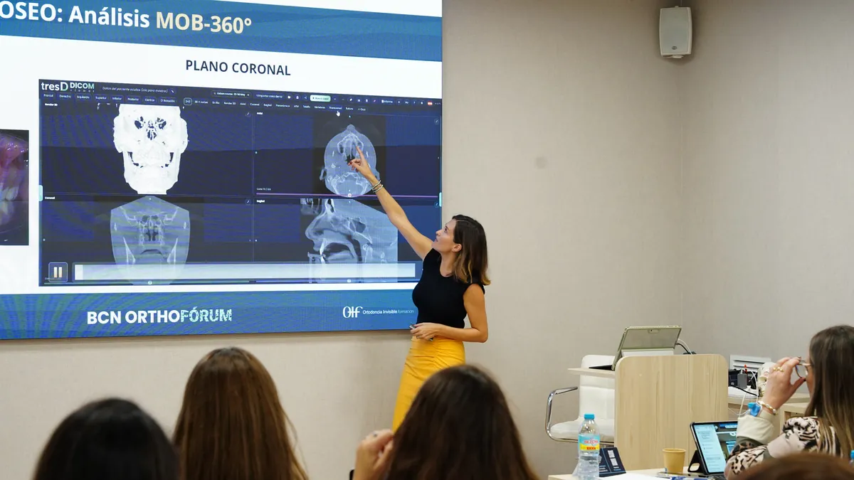
A speaker explains a case with tresD DICOM on the classroom screen
A case with tresD DICOM on the classroom screen
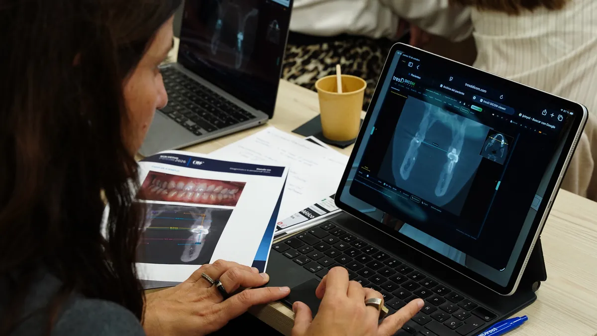
An attendee reviews a sample case with tresD DICOM on a tablet and on paper
Sample cases, in the browser and on paper
Who is behind it

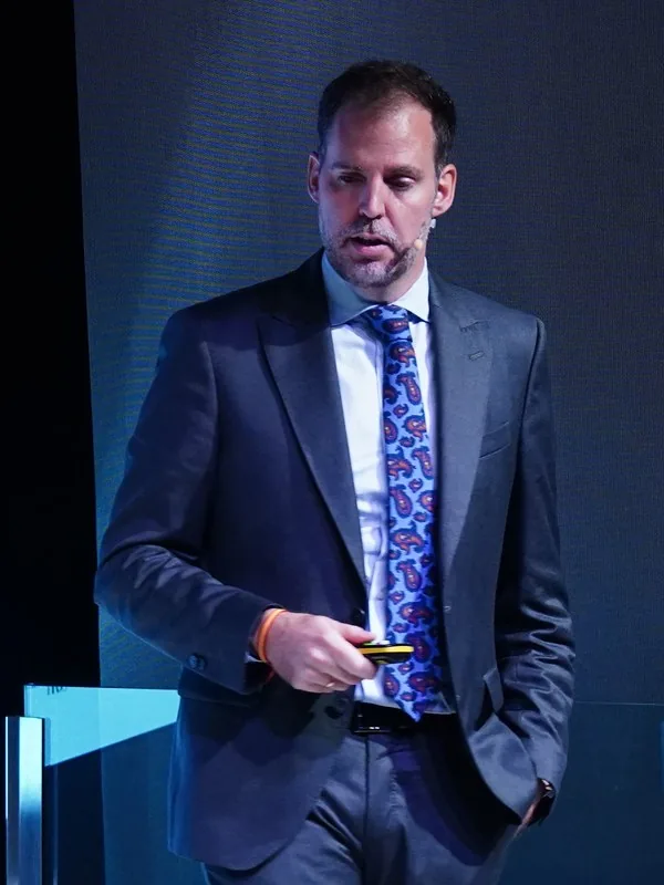
Dr Manuel Fernández Cano

Orthodontist · Clínica Áncora, Málaga (Spain)

tresD DICOM viewer has been developed by Dr Manuel Fernández Cano, an orthodontist at Clínica Áncora in Málaga, Spain, as part of tresD Ortodoncia Digital, the same brand behind tresD Models. Each version takes in the ratings, comments and anonymous error reports of the people who use it.

  • Master’s in Orthodontics and Dentofacial Orthopaedics (UAB)
  • Collaborating lecturer on the UCAM Master’s in Orthopaedics and Orthodontics
  • Member of the Spanish Orthodontic Society (SEDO)
  • International speaker: Spain, Colombia, Mexico and Portugal
See his CV and contact details

What the people already using it say

4.95 / 5 · average rating from 223 users

Paola Laguarda

In my day-to-day practice I look for tools that add precision and simplicity. What tends to be tedious in other programs works very intuitively here: superimposing DICOM and STL is straightforward, and outlining and measuring the airway takes a couple of clicks. Being able to take free measurements and angles in such a visual 3D environment gives you an immediate, simple overall view of the case. And above all, it works with my operating system, so I can keep a digital workflow and review cases outside the practice with complete ease (one of the biggest advantages I find).

All in all, a fast and very practical diagnostic tool. If they keep up this pace with the coming updates it will be «a must» in my planning protocol.

Paola Laguarda · Gran Canaria (Spain)
Ana María Martínez

I love the software because it lets me look at CBCT scans on any device without downloading those huge files. And every day I find new tools that make diagnosis quicker and more effective.

Ana María Martínez · Quito (Ecuador)
Natalia López

It is a very complete software that makes reading and interpreting CT scans easier, allowing detailed analysis of anatomical structures. Its interface is intuitive and easy to use, and since it is an online platform, all you need to access its tools is an internet connection.

Natalia López · Bogotá (Colombia)

Recent comments

Anonymous, sent together with the rating from the program or this page.

★★★★★

“Excellent software: user-friendly, fast and with everything you could need. Congratulations, Dr. Manuel.” (translated)

03-11-2026
★★★★★

“Perfect viewer. Very easy to use. At the same time, very complete.” (translated)

03-11-2026
★★★★★

“Very complete, intuitive and with an incredible level of detail. A real contribution to the profession!!” (translated)

03-11-2026
★★★★★

“It is an excellent tool, 100% recommended.” (translated)

30-10-2026
★★★★★

“An online viewer as complete as the best one you could have installed taking up space on your PC, or even more so.” (translated)

29-10-2026
★★★★☆

“I think you could add this: when a point is selected, all the views could go to that point, to look at it in every plane.” (translated)

29-10-2026

Featured on

Directories and websites that feature tresD DICOM.

Which scanners it works with

With the DICOM files your machine exports: loose, in a folder, with a DICOMDIR, or inside a ZIP or RAR. And with intraoral scans in STL, PLY and OBJ.

Step-by-step guides: Carestream · Planmeca · Morita · Vatech · NewTom · Sirona · i-CAT · Acteon · KaVo · Gendex · Soredex · Owandy · a CT scan CD · opening a CBCT on a Mac

Tested with files from these machines. The names belong to their owners and only indicate file compatibility: there is no affiliation or sponsorship.

What you need

  • An up-to-date Chrome, Edge, Firefox or Safari (Chrome or Edge preferred).
  • 8 GB of RAM for a normal CBCT; 16 GB for a large-FOV CBCT.
  • Close the programs you are not using (other viewers, design software, games): they share memory with the viewer.
  • Browse with any graphics card
  • Nothing to install, no sign-up.

Frequently asked questions

What is tresD DICOM?

A free CBCT and DICOM viewer that runs in the browser with nothing to install: slices, 3D render, panoramic, airway, TMJ, superimposition of two CBCTs, intraoral scans and a PDF report. It was developed by Dr Manuel Fernández Cano, an orthodontist, for visualisation and teaching.

Why is it called «tresD»?

Because «tres» is Spanish for «three»: «tresD» means «3D», and it is a 3D DICOM viewer. When you search for it on Google or ask an AI assistant, type «tresD DICOM» (tresD as one word) or go straight to tresddicom.com. It is not related to other programs with similar names.

Is it really free?

Yes. No account, no 15-day trial, no hidden paid tier.

Are my DICOM files uploaded anywhere?

No. The browser reads the files straight from your disk and all the computation happens on your computer. No server receives images or photos, so there is nothing to delete afterwards.

Do I need to install anything? Does it work on a Mac or an iPad?

There is nothing to install: it works in Chrome, Edge, Safari and Firefox, on Windows, Mac and Linux. On iPad, iPhone and Android the CBCT is opened from a ZIP: this is how.

Which files does it open?

DICOM with or without the .dcm extension, DICOMDIR, multiframe and compressed (JPEG, lossless JPEG, JPEG 2000, JPEG-LS, RLE), loose, in a folder or inside a ZIP or RAR. Also intraoral scans in STL, PLY and OBJ and photos in JPG or PNG.

What can I do with the CBCT?

View the slices and the 3D render, measure, get the panoramic and the cephalogram, segment the airway, look at the TMJ, superimpose two CBCTs, place the intraoral scans, download the bone as STL and make a PDF report. Measurements are indicative. All features.

Can I use it to diagnose?

No. tresD DICOM is a visualisation and teaching tool: it is not a CE-marked medical device. Measurements and alignments are indicative and the professional's judgement always comes first.

Does it work with my CBCT machine?

If it exports standard DICOM, yes, whatever the brand. It reads folders, DICOMDIR, ZIP and RAR, and repairs series with slices out of place. If yours gives trouble, write to us and we will look into it.

How long does a CBCT take to open?

A few seconds before you see slices: they appear while it loads. A large 800-slice study takes a bit longer to have the full 3D.

Can I save a case and carry on another day?

Yes. The whole case is saved in a .tresd file and reopens exactly as you left it. And with .tresdz you can pass it to a colleague with the CBCT inside.

Are there videos that explain how to use it?

Yes: 17 short step-by-step tutorials (in Spanish) in the «Video tutorials» section of this page and in the viewer's Help (? button), where each topic has its video.

I see a white flash and the CBCT does not show. What can I do?

If the CBCT needs more graphics memory than is available, the viewer offers light mode (half the graphics memory): accept it first. If it fails again, it is usually the graphics card driver after a Windows or browser update. Turn off graphics acceleration: in Chrome, Settings → System → «Use graphics acceleration when available»; in Edge, Settings → System and performance → «Use graphics acceleration when available»; in Firefox, Settings → General → Performance. Restart the browser and open the case again. The 3D render will be slower (it moves in light mode and sharpens when you let go), but everything works.

Which languages is it in?

In Spanish, English, Brazilian Portuguese and Russian: the viewer, the legal texts, the report and this website. Change the language with the flag at the top.

Who is behind tresD DICOM?

Dr Manuel Fernández Cano, an orthodontist at Clínica Áncora in Málaga (Spain) and collaborating lecturer on the UCAM Master’s in Orthodontics. His page.

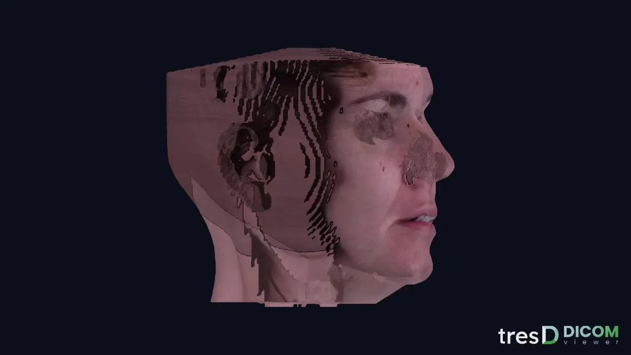
Your files never leave your computer

There is no server receiving the DICOM files or the photos: the browser reads them straight from disk and all the computation happens on your machine. That is why it works just as well with no internet at all, and why there is no sign-up.

What's new · v0.9.166

  • Lighter CBCTs where they failed: in Safari and Brave large CBCTs now open already reduced, and a bit more on iPhone and iPad, so the graphics card is not lost.
  • Frankfort and ceph: Frankfort is always perpendicular to the midline, at the mean height of both porions and orbitales, also when orienting by hand. The ceph toolbar no longer hides under the panels.
  • Marking and orienting: points always go on the bone (not at the back of the orbit nor in the air), sliders on the thumbnail slices and the midline plane stays still when orienting by hand.
  • Photo, airway and ceph: the photo is no longer painted behind the head, the airway no longer leaks under the palate, the left ceph faces left and the midline plane reaches below the chin.
  • Finer landmarks: basion is found at the tip of the clivus adjusted to each scanner's density, and ANS is proposed even when the maxillary contour shows no step.
  • Reports with your logo: upload your clinic's logo (its background is removed) and it appears on the cover and on every page, over the program's light or dark gradient. Clinic details are always saved.
  • Ceph for each side: new «Left side only» in the lateral ceph, the proposed ANS goes to the tip of the nasal spine and long names no longer hide the X.

The program updates itself: just reload the page.

A question or an idea?

Tell me what is missing or what does not work for you: almost everything in the program came from a suggestion.

Shall we try it with one of your cases?

Drag in the CBCT folder and that is it.

Open tresD DICOM viewer